

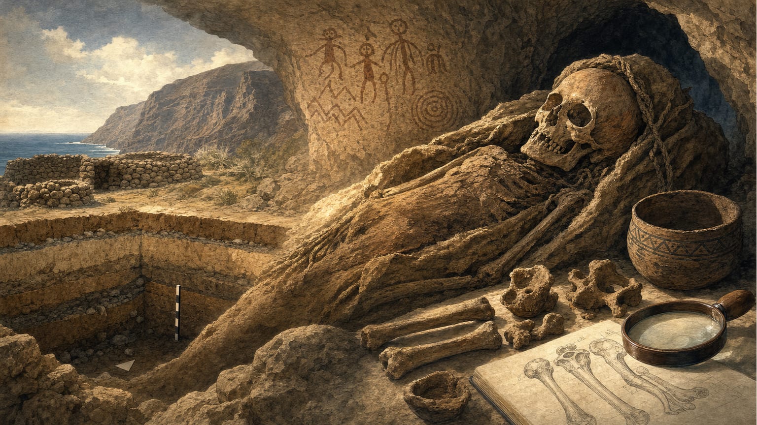
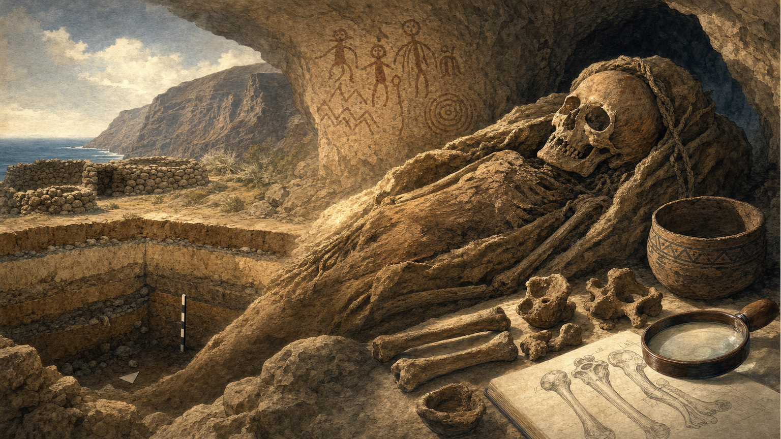
Bone paleopathology, the study of diseases in ancient skeletal remains, has been a central focus in the analysis of Guanche mummies from Tenerife within the CRONOS Project, launched by the Archaeological and Ethnographic Museum of Tenerife in 1989[1]. This project has enabled the identification and quantification of various pathologies in the island’s pre-Hispanic indigenous population, shedding light on their health and living conditions.
One of the most significant findings is the high frequency of degenerative joint disease (DJD), also known as osteoarthritis. According to the data, between 30% and 35% of the specimens studied showed signs of DJD in one or more joints[1]. The elbow joint was the most affected (30%), followed by the shoulder and knee (20% each), and the hip with a slightly lower percentage. It was observed that men tended to have more knee and shoulder problems, while women had a higher incidence of coxarthrosis. Additionally, sacroiliac DJD mainly affected women (10%), and other joints such as the wrist (4%) and ankle (2%) were less frequently involved[1].
The high prevalence of these joint diseases has been linked to a vigorous lifestyle with intense physical activity, especially from the third decade of life, as evidenced by the high incidence of spondylosis (25%) from ages 25-30[1]. Researchers suggest that activities such as gathering and stone-throwing may have contributed to these injuries, supported by the presence of exostoses in the ulnar notch of the ulna.
Other joint pathologies included ankylosing spondylitis, detected in about 1.2% of cases, mainly affecting men—a frequency similar to that seen in the current island population[1]. Only one possible case of rheumatoid arthritis was identified, though the diagnosis remains uncertain.
Bone trauma was also studied. Cranial fractures had a global incidence of 7% in the sample (1000 skulls), with a 4:1 ratio between the south and north of the island. Most of these injuries were related to violence, especially in young men aged 18 to 35. Depressed and irregular fractures located in the anterior and lateral parts of the skull were associated with the impact of lithic objects[1]. Postcranial fractures were less frequent (less than 1%), mostly affecting the upper limb.
Regarding tumors, no primary or metastatic malignant tumors were found, but benign osteomas were observed in the cranial vault (2-4%) and solitary exostoses near the growth cartilage of long bones, with a higher incidence in men[1].
As for metabolic disorders, senile osteoporosis was the most common alteration, especially in women, while diseases such as rickets or scurvy were not detected in the pre-Hispanic population studied[1].
Finally, spina bifida occulta stood out as the most frequent congenital malformation, reaching over 50% in some series and with a global average of 35%, higher in men. The authors suggest that endogamy and biological ties with the North African Berber population could explain this high frequency[1].